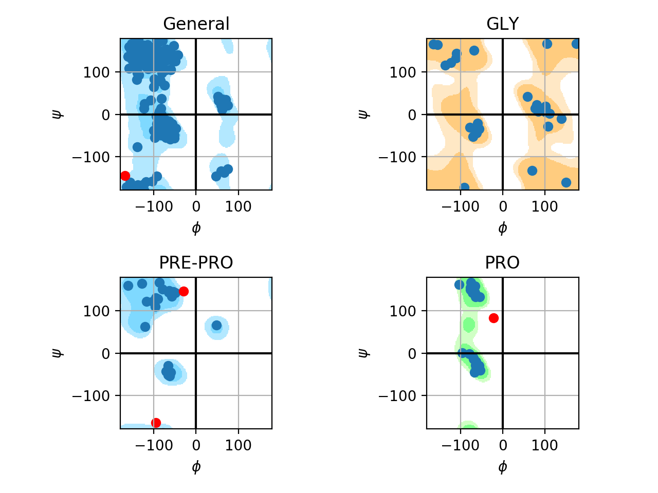
Ramachandran plot

| Poses (avg) | Affinity (wild) | Affinity (mutant) | ΔAffinity | ΔAffinity expected | Status |
|---|---|---|---|---|---|
| 1 | -7.40 | -7.20 | 0.2 | ΔAffinity < 0 | x |
| 1-3 | -6.93 | -6.77 | 0.16 | ΔAffinity < 0 | x |
| 1-10 | -6.54 | -6.36 | 0.18 | ΔAffinity < 0 | x |
| Poses (avg) | Affinity (wild) | Affinity (mutant) | ΔAffinity | ΔAffinity expected | Status |
|---|---|---|---|---|---|
| 1 | -7.40 | -5.60 | 1.8 | ΔAffinity > 0 | ✓ |
| 1-3 | -6.93 | -5.27 | 1.66 | ΔAffinity > 0 | ✓ |
| 1-10 | -5.16 | -5.07 | 0.09 | ΔAffinity > 0 | ✓ |
| Poses (avg) | Affinity (wild) | Affinity (mutant) | ΔAffinity | ΔAffinity expected | Status |
|---|---|---|---|---|---|
| 1 | -7.40 | -7.17 | 0.23 | ΔAffinity < 0 | x |
| 1-3 | -6.93 | -6.86 | 0.069999999999999 | ΔAffinity < 0 | x |
| 1-10 | -6.54 | -6.52 | 0.02 | ΔAffinity < 0 | x |
| Poses (avg) | Affinity (wild) | Affinity (mutant) | ΔAffinity | ΔAffinity expected | Status |
|---|---|---|---|---|---|
| 1 | -7.40 | -5.67 | 1.73 | ΔAffinity > 0 | ✓ |
| 1-3 | -6.93 | -5.47 | 1.46 | ΔAffinity > 0 | ✓ |
| 1-10 | -5.16 | -5.23 | -0.07 | ΔAffinity > 0 | x |
Three-dimensional structures were modeled by homology using MODELLER. Sequences were collected in UniProt. We searched for the E.C. number of beta-glucosidases (3.2.1.21) and collected sequences of family GH1 (~3000). For each sequence, we selected the structure of highest identity using BLAST against the PDB database to be used as a template. Sequences with identity lower than 25% were discarded. 100 models were constructed for each sequence. The best model was selected using the lowest value of the DOPE score. Mutants were modeled using MODELLER's mutation script. The mutations were based on beneficial mutations described in the literature.
PDB models were minimized using AMBER tools. We performed 1000 steps of minimization in the vacuum (parameters: imin=1, maxcyc=1000, ncyc=750, cut=10.0, ntb=0, igb=0).
We performed multiple alignment using Clustal Omega (default parameters). We used Python scripts to detect conservation.
We performed docking of glucose (product) and cellobiose (substrate) for wild and mutant beta-glucosidases using AutoDock Vina (parameters: exhaustiveness = 20; num_modes = 10). The box center was defined using the median coordinate of the last atom of the two catalytic glutamates. The experiment was performed in triplicate. We collected ten poses for each docking and analyzed the average of affinity energies predicted by the software for (i) pose 1, (ii) poses 1-3, and (iii) poses 1-10.